Automated enhancement of data quality

The goal of this research area is the development of techniques that enhance the quality of routine clinical data, which is often inferior due to, e.g., the tight schedule of clinical routine, limited time in emergency settings, and a lack of patient cooperation during acquisition. Imaging data with a reduced quality are frequently characterized by a higher noise level, motion artefacts, and missing components, as for instance a missing T2-weighted scan in a typical clinical scanning protocol for the acquisition of structural brain data. Hence, techniques for noise reduction, motion correction, and image synthesis, i.e. the imputation of missing contrasts/sequences, are required.
In collaboration with the Department of Biomedical Magnetic Resonance (Oliver Speck) at the Otto-von-Guericke University Magdeburg, we develop algorithms for noise reduction and motion correction of Magnetic Resonance Imaging (MRI) brain scans. In particular, denoising during image reconstruction and deep learning approaches to motion correction are investigated. The level of quality enhancement is then, determined using an automated digital analysis.
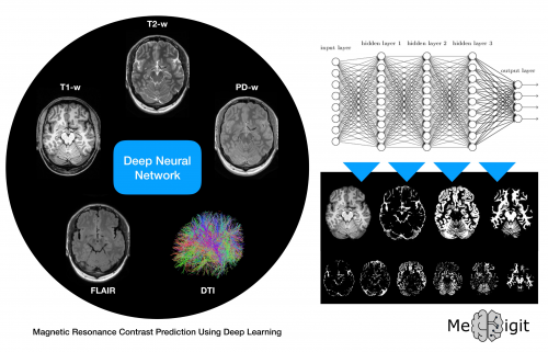
Image synthesis is an active research field gaining pace with recent developments in deep learning that allow for MRI contrast imputation based on available measured data. In this research area, we develop a framework that facilitates the prediction of an arbitrary contrast based on the given ones. For this purpose, we also explore possibilities to enhance the prediction through data from different imaging modalities, such as, CT (Computed Tomography), PET (Positron Emission Tomography), SPECT (Single-photon emission computed tomography), NIRS (Near-infrared spectroscopy), EEG (Electroencephalography), etc. The uncertainty introduced by data imputation must be quantified and considered during feature extraction and biomarker discovery.
Image data acquired for research purposes are of higher quality as compared to the routine data since longer measurement times can be invested and subjects in general collaborate well. High-resolution MRI data can be acquired using high field strengths (7T). Associations between low- and high-resolution data of the same subject can be learnt and employed for predicting details in the former (super-resolution). Along these lines, we research methods for enhancing the resolution in routine clinical data.


